Plantar Foot Muscles Mri - Nodules or masses of plantar fibromatosis are typically located in the middle to the medial aspect of the plantar arch and may extend to involve the skin or deep structures of the foot.
Plantar Foot Muscles Mri - Nodules or masses of plantar fibromatosis are typically located in the middle to the medial aspect of the plantar arch and may extend to involve the skin or deep structures of the foot.. The plantar fascia is a multilayered, fibrous aponeurosis with medial, central, and lateral components (, 1).the term plantar fascia typically refers to the large central component, which originates from the medial calcaneal tuberosity and extends anteriorly, adhering to the underlying flexor digitorum brevis (fdb) muscle. At mr imaging, the course of the plantar tendons is optimally visualized with dedicated imaging of the midfoot and forefoot. Medial sides of metatarsals of toes iii to v insertion: Muscles that move the foot and toes. Please come back soon to see the finished work!
Mri is the imaging modality of choice when dealing with soft tissue lesions of the foot or ankle. The origins of the lumbrical muscles are located at the distal end of the quadratus plantae muscle. The plantar fascia is a multilayered, fibrous aponeurosis with medial, central, and lateral components (, 1).the term plantar fascia typically refers to the large central component, which originates from the medial calcaneal tuberosity and extends anteriorly, adhering to the underlying flexor digitorum brevis (fdb) muscle. The medial plantar vein lies between the abductor hallucis and the flexor hallucis brevis muscles. Smaller terminal division of the tibial nerve course:
Calcaneal Spurs Physiopedia from www.physio-pedia.com The quadratus plantae muscle runs immediately deep to it. Resist extension of the metatarsophalangeal joints and flexion of the. Please come back soon to see the finished work! These include plantar fibromatosis, haemangioma, lipoma, pvns/gct tendon sheath and synovial chondromatosis. The abductor digiti minimi muscle is located on the lateral side of the foot. Mri has surpassed nuclear medicine imaging due to the greater specificity of mri and its ability to delineate osseous anatomy as well as discrete abscesses and sinus tracts diagnostic of infection. At about the midsole, it splits into five. Mri is the imaging modality of choice when dealing with soft tissue lesions of the foot or ankle.
The plantar fascia is a fibrous aponeurosis that arises along the medial calcaneal tuberosity.
Please come back soon to see the finished work! The plantar fascia is a fibrous aponeurosis that arises along the medial calcaneal tuberosity. Findings on conventional arthrography and mr imaging. The plantaris muscle is one of the calf muscles in the superficial posterior compartment of the leg. Magnetic resonance images of the foot may be digitized to quantify muscle architecture. Plantar plate of the foot: Lesions may be symptomatic because of a mass effect or invasion of adjacent muscles or neurovascular structures. At mr imaging, the course of the plantar tendons is optimally visualized with dedicated imaging of the midfoot and forefoot. Mri features of palmar and plantar fibromatosis. The quadratus plantae muscle runs immediately deep to it. Chronic plantar fasciitis may be accompanied by muscle atrophy of plantar intrinsic foot muscles and tibialis posterior compromising the dynamic support of the foot prolonging the injury. 23,25 mri at the level of the malleolus demonstrates the muscle as. It contributes to the surface anatomy of the medial sole of the foot and is easy to palpate.
The plantar fascia is a multilayered, fibrous aponeurosis with medial, central, and lateral components (, 1).the term plantar fascia typically refers to the large central component, which originates from the medial calcaneal tuberosity and extends anteriorly, adhering to the underlying flexor digitorum brevis (fdb) muscle. Extensor hoods and bases of proximal phalanges of toes iii to v action: The plantar arch, plantar metatarsal veins and the medial and lateral plantar veins lie deep to plantar muscle groups. 23 it can originate as a separate muscle from the fibula or from the peroneus brevis or longus muscles and inserts onto the peroneal tubercle or retrotrochlear eminence of the calcaneus. Muscle hernia is optimally visualized with us, but dynamic mr imaging with the foot in plantar flexion and dorsiflexion can also be used.
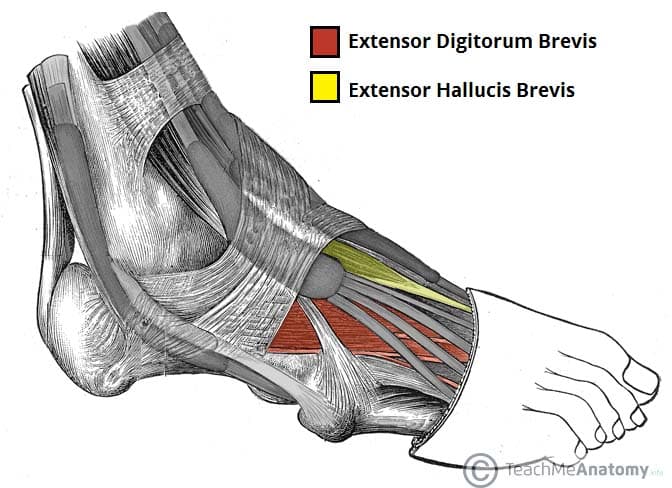
Muscles Of The Foot Dorsal Plantar Teachmeanatomy from teachmeanatomy.info Lesions may be symptomatic because of a mass effect or invasion of adjacent muscles or neurovascular structures. 23 it can originate as a separate muscle from the fibula or from the peroneus brevis or longus muscles and inserts onto the peroneal tubercle or retrotrochlear eminence of the calcaneus. In addition, an image of all the muscles of the back and plantar part of the foot, all tendons and tendon ligaments, blood vessels and nerves are obtained. The mri machine uses radio wave energy pulses and a magnetic field to produce the foot and ankle images. Plantar fasciitis refers to inflammation of the plantar fascia of the foot. To describe changes in activation of the intrinsic plantar foot muscles after 4 exercises as measured with t2 magnetic resonance imaging (mri). A case report and review of anatomy. Heel spurs and plantar fasciitis are not the same thing, and heel spurs do not cause plantar fasciitis.
9 yao l, do hm, cracchiolo a, et al.
Are plantar fasciitis and heels spurs the same thing? Familiarity with the normal anatomy of the plantar tendons and its appearance at magnetic resonance (mr) imaging and ultrasonography (us) is essential for recognizing plantar tendon disorders. Chronic plantar fasciitis may be accompanied by muscle atrophy of plantar intrinsic foot muscles and tibialis posterior compromising the dynamic support of the foot prolonging the injury. 2 from the calcaneus, the plantar fascia divides into the medial, central, and lateral components (figure 1). These include plantar fibromatosis, haemangioma, lipoma, pvns/gct tendon sheath and synovial chondromatosis. Mri findings of acute turf toe: 10 foot and ankle craig r. The plantar fascia is a multilayered, fibrous aponeurosis with medial, central, and lateral components (, 1).the term plantar fascia typically refers to the large central component, which originates from the medial calcaneal tuberosity and extends anteriorly, adhering to the underlying flexor digitorum brevis (fdb) muscle. Medial sides of metatarsals of toes iii to v insertion: 23,25 mri at the level of the malleolus demonstrates the muscle as. A case report and review of anatomy. The interosseous muscles of the fourth interspace are usually supplied by a branch from the superficial ramus of the lateral plantar interosseous, plantar the plantar interosseous muscle arises from the proximal third of the medial plantar surface of the shaft, from the base of the metatarsal on which it lies, and from the fascial expansions of. Adduction of toes iii to v at metatarsophalangeal joints;
In the past, these bone spurs were often blamed for heel pain and removed surgically. Chronic plantar fasciitis may be accompanied by muscle atrophy of plantar intrinsic foot muscles and tibialis posterior compromising the dynamic support of the foot prolonging the injury. In addition, an image of all the muscles of the back and plantar part of the foot, all tendons and tendon ligaments, blood vessels and nerves are obtained. The medial plantar vein lies between the abductor hallucis and the flexor hallucis brevis muscles. Plantar fasciitis refers to inflammation of the plantar fascia of the foot.
Master Knot Of Henry Revisited A Radiologist S Perspective On Mri Clinical Radiology from els-jbs-prod-cdn.jbs.elsevierhealth.com In the past, these bone spurs were often blamed for heel pain and removed surgically. Chronic plantar fasciitis may be accompanied by muscle atrophy of plantar intrinsic foot muscles and tibialis posterior compromising the dynamic support of the foot prolonging the injury. The lateral plantar vein lies between the flexor digitorum brevis muscle and quadratus plantae. The interosseous muscles of the fourth interspace are usually supplied by a branch from the superficial ramus of the lateral plantar interosseous, plantar the plantar interosseous muscle arises from the proximal third of the medial plantar surface of the shaft, from the base of the metatarsal on which it lies, and from the fascial expansions of. The origins of the lumbrical muscles are located at the distal end of the quadratus plantae muscle. Findings on conventional arthrography and mr imaging. Mri has surpassed nuclear medicine imaging due to the greater specificity of mri and its ability to delineate osseous anatomy as well as discrete abscesses and sinus tracts diagnostic of infection. Familiarity with the normal anatomy of the plantar tendons and its appearance at magnetic resonance (mr) imaging and ultrasonography (us) is essential for recognizing plantar tendon disorders.
Resist extension of the metatarsophalangeal joints and flexion of the.
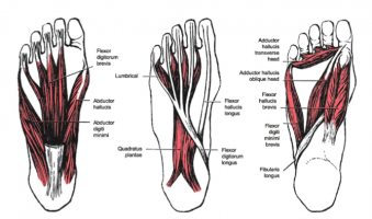
23 it can originate as a separate muscle from the fibula or from the peroneus brevis or longus muscles and inserts onto the peroneal tubercle or retrotrochlear eminence of the calcaneus. The medial plantar vein lies between the abductor hallucis and the flexor hallucis brevis muscles. It attaches to the lateral base of the proximal phalanx of the 5th digit. It contributes to the surface anatomy of the medial sole of the foot and is easy to palpate. It is homologous with the abductor digiti minimi of the hand. Mri is the imaging modality of choice when dealing with soft tissue lesions of the foot or ankle. 6 mri is commonly ordered in the diabetic patient to rule out infection in the presence of an ulcer, to evaluate the severity of charcot arthropathy. Muscles that move the foot and toes. 2 from the calcaneus, the plantar fascia divides into the medial, central, and lateral components (figure 1). The muscles lying within the medial group form a bulge referred to as the 'ball' of the big toe. Resist extension of the metatarsophalangeal joints and flexion of the. Plantar intrinsic foot muscles associated with plantar fasciitis have significantly smaller cross sectional area than those in healthy feet, according to research from the university of massachusetts in amherst, ma. The three groups of plantar foot muscles are(14):
Certain soft tissue tumours are identifiably benign because of their signal characteristics, morphology and/or location foot muscles mri. The plantar fascia is a multilayered, fibrous aponeurosis with medial, central, and lateral components (, 1).the term plantar fascia typically refers to the large central component, which originates from the medial calcaneal tuberosity and extends anteriorly, adhering to the underlying flexor digitorum brevis (fdb) muscle.


Posting Komentar
0 Komentar